Respiratory cycle
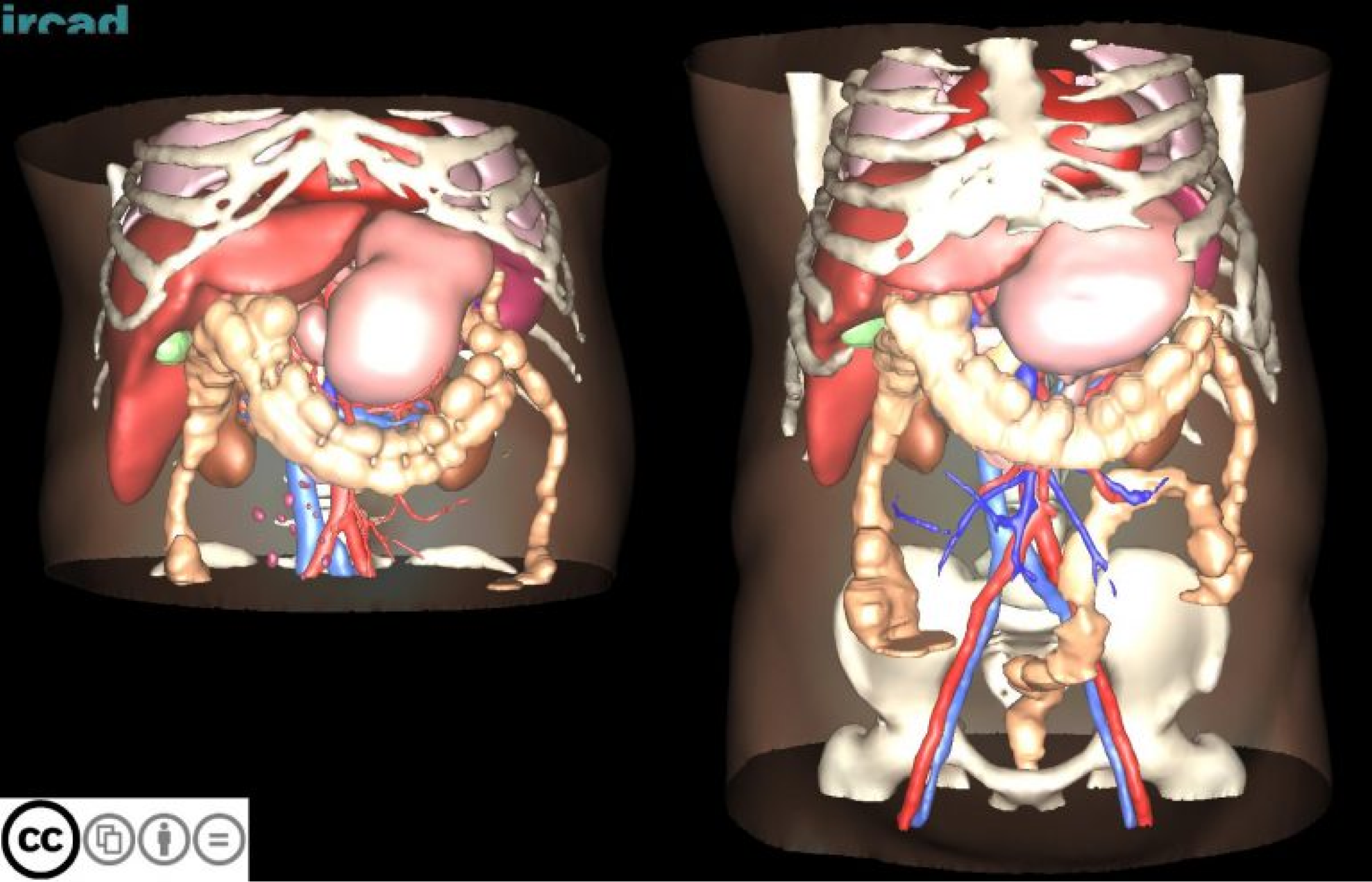
The 3D-IRCADb-02 database is composed of two anonymized 3D CT-scans.
Both folders corresponding to those two acquisitions can be downloaded individually or conjointly.
The first one has been realized during the arterial phase in inhaled position, whereas the second one
has been realized during the portal phase in exhaled position. The patient has a hepatic focal nodular
hyperplasia in segment VII according to Couinaud’s description.
These folders are called “3D-IRCADb2.number” (the number varying between 1 and 2).
Each “3D-IRCADb-2-number”
folder contains 4 sub-folders called “PATIENT_DICOM”, “LABELLED_DICOM”, “MASKS_DICOM” and “MESHES_VTK”.
These folders respectively contain the anonymized patient image in DICOM format, the labelled image corresponding
to the various zones of interest segmented in DICOM format, a new set of sub-folders corresponding to the names
of the various segmented zones of interest containing the DICOM image of each mask, and finally, all the files
corresponding to surface meshes of the various zones of interest in VTK format.
Size: 116Mb
Description of the 2 cases of 3D-IRCADb-02


This work is licensed under a Creative Commons Attribution-NonCommercial-NoDerivatives 4.0 International License.
For reference, please cite our publication below. For any further information, please contact us.
Soler, L., A. Hostettler, V. Agnus, A. Charnoz, J. Fasquel, J. Moreau, A. Osswald, M. Bouhadjar, and J. Marescaux. “3D image reconstruction for comparison of algorithm database: A patient specific anatomical and medical image database.” IRCAD, Strasbourg, France, Tech. Rep (2010)