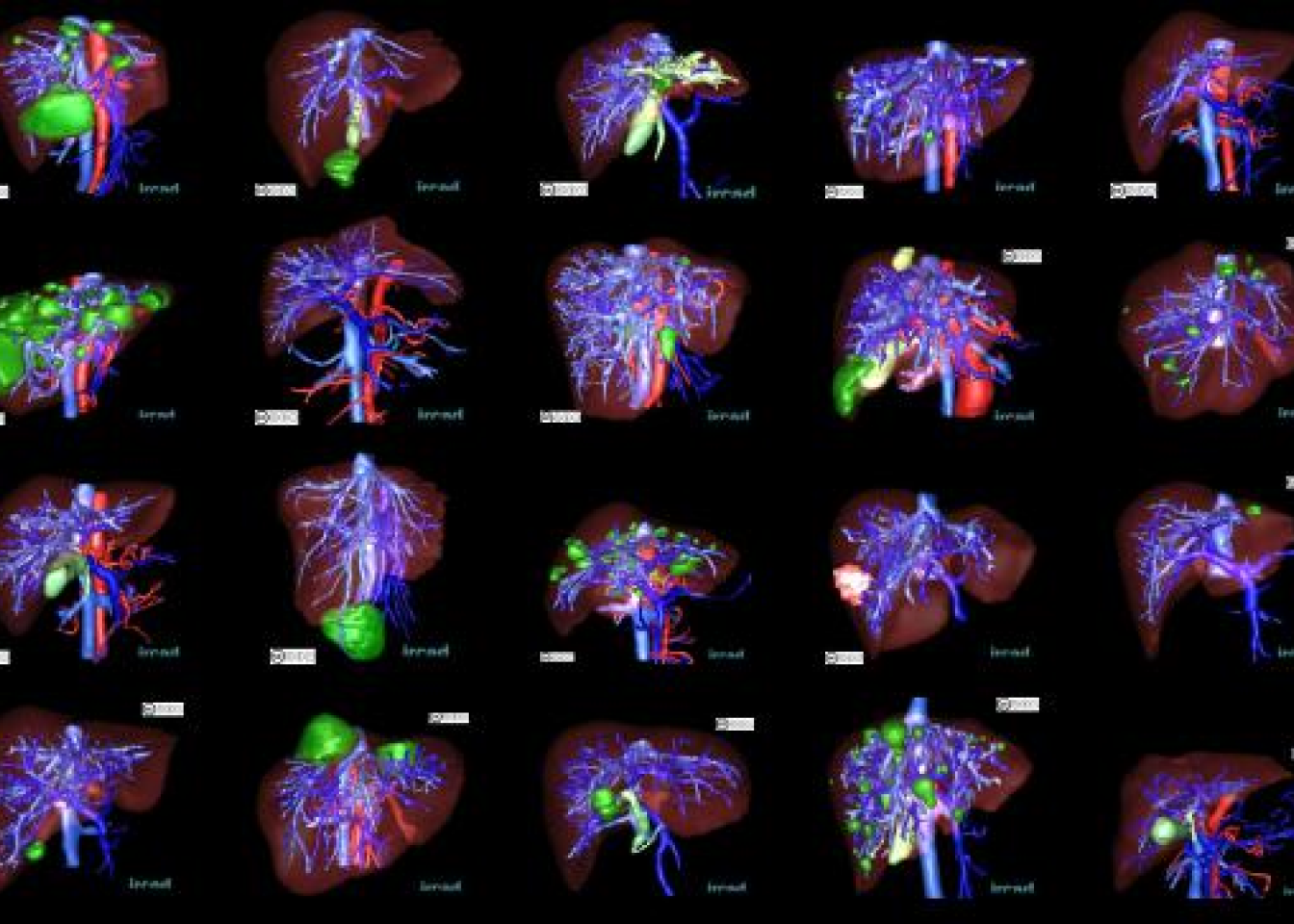
Liver segmentation 3D-IRCADb-01
The 3D-IRCADb-01 database is composed of the 3D CT-scans of 10 women and 10 men
with hepatic tumours in 75% of cases. The 20 folders correspond to 20 different patients, which can be
downloaded individually or conjointly. The table below provides information on the image, such as liver size
(width, depth, height) or the location of tumours according to Couninaud’s segmentation. It also indicates the
major difficulties liver segmentation software may encounter due to the contact with neighbouring organs, an
atypical shape or density of the liver, or even artefacts in the image.
These folders are called “3D-IRCADb-1-number” (the number varying between 01 and 20).. These folders are called “3D-IRCADb-01-number” (the number varying between 01 and 20). Each “3D-IRCADb-01-number” folder contains 4 sub-folders called “PATIENT_DICOM”, “LABELLED_DICOM”, “MASKS_DICOM” and “MESHES_VTK”. These folders respectively contain the anonymized patient image in DICOM format, the labelled image corresponding to the various zones of interest segmented in DICOM format, a new set of sub-folders corresponding to the names of the various segmented zones of interest containing the DICOM image of each mask, and finally, all the files corresponding to surface meshes of the various zones of interest in VTK format.
Size: 782Mb
Description of the 20 cases of 3D-IRCADb-01
Overview
N°
Sex
Year of birth
Voxel size (mm)
Image size (pixels)
Liver size (cm)
Liver Average density
Liver pathologies
Segmentation drawbacks
Download

1
F
1944
0,57
0,57
1,6
512
512
129
18,3
15,1
14,1
111
7 tumours in III, IV/V, VII, VIII
stomach, pancreas, duodenum
Size 37Mb


This work is licensed under a Creative Commons Attribution-NonCommercial-NoDerivatives 4.0 International License.
For reference, please cite our publication below. For any further information, please contact us.
Soler, L., A. Hostettler, V. Agnus, A. Charnoz, J. Fasquel, J. Moreau, A. Osswald, M. Bouhadjar, and J. Marescaux. “3D image reconstruction for comparison of algorithm database: A patient specific anatomical and medical image database.” IRCAD, Strasbourg, France, Tech. Rep (2010)